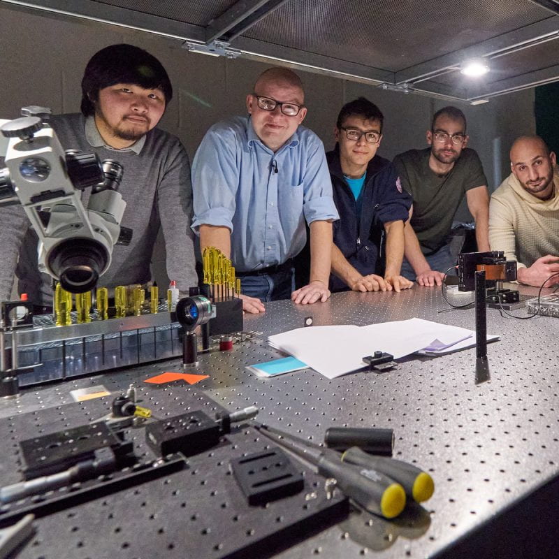
Imagine the brain as a universe inside each of us, full of mysterious energies that interact with one another, creating galaxies of thought and movement. Now imagine we are able to map that universe, explore it, understand it, and ultimately fix it when it goes wrong. Just like our exploration of outer space, says Chris Xu, Applied and Engineering Physics, our exploration of the brain starts with the development of new technologies that will allow us to uncover its secrets.
“Understanding how the brain works is the Holy Grail in science in many ways,” Xu says. He explains the current dilemma in brain studies: On the one hand, neuroscientists have a great deal of knowledge of how one neuron works and have observed a handful of neurons interacting in the lab. Scientists studying human behavior, on the other hand, have set up MRI experiments mapping areas of the brain that react to certain stimuli. For instance, they’ve shown photos of babies to adults and recorded which part of the brain responded. These areas identified through MRIs are typically about a million neurons in size. “But what happens between a handful of neurons and a million neurons?” Xu asks. “We don’t know. We don’t have the tools we need to study the phenomenon in between.”
To See Inside the Brain
Xu wants to fill that technology gap. As part of the White House’s Brain Research through Advancing Innovative Neurotechnologies (BRAIN) Initiative, he and his lab are working on a new type of optical imaging that will allow scientists to observe neurons functioning inside a living brain at a high temporal and high spatial resolution. Currently, they are testing the new technology on mouse models.
“We have made a lot of progress in seeing deep inside the living mouse brain with high resolution,” Xu says. He developed a groundbreaking microscope that uses lasers and fluorescent tags at the cellular level to register brain activity. “It’s noninvasive, so we can look at the brain of a live mouse without damaging it. A mouse brain is about eight millimeters thick, and we can see to a depth of one to two millimeters, at this point. This is much deeper than anyone had ever seen previously.”
Xu’s goal is to penetrate ever deeper into the layers of the brain, making large-scale volumetric recordings of brain activity. “Once we can see a large volume of neurons, we can measure the neuronal activity of the whole functional units,” he says. “If we can understand how functional units of the brain work—for instance, the visual cortex or the motor cortex—we can put those units together to eventually have a better understanding of the whole brain.”
Telecom, Fiber Optic, and Imaging Techniques—Thinking Outside the Box
Xu and his lab are able to develop this state-of-the-art imaging tool because of the broad range of expertise he brings to the problem. After receiving his PhD training in imaging at Cornell, he went on to work in the telecommunications industry for five and a half years before returning to Cornell as a professor in the early 2000s. Those years working on the industry side taught him all about fiber optics and telecom techniques, he says, which he has since put to use on a range of academic questions, including brain imaging.
“We have a unique combination of telecom, fiber optic, and imaging techniques,” he says. “It’s a beautiful integration of these seemingly different directions. We combine this expertise with an understanding of the huge problem we have in brain science: the lack of knowledge about the functional unit level of the brain. This is what drives us to the forefront of this brain imaging effort.”
“If we can understand how functional units of the brain work—for instance, the visual cortex or the motor cortex—we can put those units together to eventually have a better understanding of the whole brain,” Xu says.
Xu’s brain-imaging microscope uses lasers created through fiber optic techniques developed in his lab. The large-scale recordings of the brain are made possible by a common modulation technique used in the telecom industry. “But it’s a technique that’s brand new to imaging and neuroscience,” he says. “Seeing that connection between telecom technology and brain imaging, that’s thinking outside the box.”
Endoscope, an Eye Inside a Body
Xu has also applied his thinking-outside-the-box skill to other medical technology, such as endoscopy. In particular he is collaborating with medical doctors at Weill Cornell Medicine and Mount Sinai Hospital to develop miniaturized microscopes that can be inserted inside the human body, allowing doctors to observe tissues at the cellular level.
“Right now endoscopy is like putting your eye inside a body,” Xu says. “That’s the level of visualization you have. You can see discoloration of tissue and maybe inflammation. If you’re looking for an early stage cancer, the next step is to get your scalpel and take a piece of tissue out and then send it to a lab where they’ll look at it under a microscope. A few days later you get the results.”
Xu and his collaborators want to eliminate the tissue biopsy and the wait. They are creating a microscope so small, similar in size to a biopsy needle, that it can be inserted through a catheter directly to the tumor site. “It’s real-time diagnosis,” Xu says. “No tissue removal.” The tiny, powerful endoscope could also help doctors avoid collateral damage, such as mistakenly cutting nerve fibers during surgery.
“We have to figure out how to maintain the capability that already exists in microscopes but shrink it to a size we can insert inside the human body,” Xu says. “This is a real engineering challenge.”
Researchers have created prototypes in the lab and tested them on live animal models. And they used their endoscope to analyze excised human specimens at Weill Cornell Medicine. The comparison of the researchers’ results with the official lab results looked very promising.
“We are getting the clinical validation of our technology, so we can prove our endoscope can be used with confidence,” Xu says. “Our goal is to translate this technology to the hospital. It will have a dramatic, immediate impact on human health and patient care.”